Swollen feet, should I be concerned?

As with any medical problem, understanding the cause or diagnosis is the first step to the right treatment. Swelling or to give it the correct medical term Oedema in the feet and ankles can have many causes. I thought I would talk about some of the more common causes.

There are two general categories of oedema seen in the feet. 1. Pitting oedema is as the name suggests, leaves a pit or indentation when the area is pressed with the finger and will be visible for a little time after you have pressed the area.


2. Non pitting oedema, (I think you can see where this is going) when you press the area with your finger it doesn’t leave an indentation.


Pitting oedema is generally caused by fluid, mainly water accumulating in the feet and ankles. Due to the effects of gravity the pitting oedema will be worse as the day progresses. Pitting oedema is commonly caused by poor circulation or the retention of excess fluids. Some common risk factors that may lead to these problems include sitting or standing in one position for too long, low protein levels, obesity, high salt intake, dehydration, heart failure, varicose veins, cirrhosis and pregnancy.  Some medications may also cause oedema in the feet and legs, some blood pressure medications, steroid therapy, hormone therapy, contraceptive pill and even some antidepressants. All of the above causes are exacerbated by warm weather when we push more blood to the skin to try to lose heat. So I definitely see more pitting oedema in the summer here in Spain.


Non pitting oedema, is most often caused by problems with Thyroid, or the lymphatic system, lymph nodes are sometimes removed as part of cancer treatment which can lead to non pitting oedema. Lipedema ( a buildup of fat under the skin ) also causes non pitting oedema.


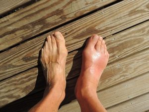
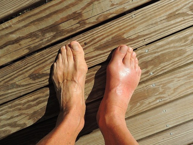
Generally speaking all of the above are seen in both feet and legs ( bilateral ) with maybe the exception of where varicose veins are particularly bad in one leg, ( pitting oedema ) or a lymph node is taken from just one side of the body ( non pitting oedema). There are some other non-pitting types of oedema which affect a single foot or leg, these would include, sprains and injuries, infection, and even deep vein thrombosis ( known as DVT or blood clot ). Although most oedema is associated with some discomfort,  injury, infection and DVT are usually associated with acute pain, redness of the skin and heat. Untreated oedema can result in foot and leg ulcers.


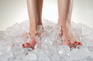
Treatment, as stated at the beginning understanding the underlying cause is the most important thing often treating the underlying cause will relieve symptoms. With single foot non pitting oedema treating the underlying condition will resolve the issue.  But treating the swelling with pitting oedema ( once the cause is established ) will fall roughly into three categories: Mild cases should resolve on its own by elevating legs and feet, generally above the the backside or level with the heart is even better. Severe cases diuretic tablets to help eliminate excess fluid via urine. Chronic cases compression stockings are required.

Cold Feet

Do you have cold feet?

If you have cold feet and the rest of you is warm, it could be a sign of poor circulation.

Particularly this time of year I seem to spend a lot of time doing vascular assessments. The colder weather often exacerbates poor circulation resulting in cold feet, colour changes, chilblains, discomfort and in some cases ulceration to the feet.

Although feeling pulses in the feet is a good initial way of assessing circulation. There are some basic observations that can also give clues to what arterial blood supply (that’s blood coming down to the feet) is like. Skin texture can be a good pointer, thin shiny skin and lack of hairs often indicates that blood supply is not very good. Capillary refill time is the time it takes for the blood to come back after it has been squeezed out, for example if you press your finger when it is raised above your heart, you will see the skin go white. Capillary refill time is the time it takes for the blood to come back to the skin, the normal is two seconds or less.

Other symptoms of poor circulation can be pain in the calf muscle when walking, this is known as claudication pain. The pain will come on after a certain distance, known as the claudication distance. What is happening here is when you walk the calf muscle requires blood and if circulation is poor after walking a while the muscle has used the available blood and the resulting pain is the muscle telling us it needs more blood. Normally people will stop walking and the pain will subside. Claudication distance will decrease with incline, so for example if your claudication distance is 150 m on the flat it may be only 50m on a hill due to the muscles having to work harder.

Although vascular assessment is about patient history, physical signs and symptoms. Doppler is a great tool. Doppler assessment is a method of listening to the pulses in the feet. Doppler uses ultrasound to to bounce off circulating red blood cells to measure blood flow. Doppler is non invasive and painless an can give a very accurate picture of what the circulation is like in the feet.

If needed or requested Vascular assessment including doppler is part of a normal appointment. So why not have your circulation checked.

Philip Mann Podiatrist/Chiropodist 686912307 philipmann@footpodiatrist.com

Clinica Alegria welcomes a new doctor

Clinica Alegria is pleased to welcome Dr. Loes Thomaes to their practice

Loes Thomaes General Practitioner (English, Dutch, Spanish, German and French speaking).

Studied medicine at “de Vrije Universiteit” in Amsterdam and completed her residency in Leiden. As a General Practitioner she worked in many practices in and around The Hague. Furtheron she has worked many years as a medical doctor in several Dutch hospitals at the Geriatric and Psychiatric departments.

In spring 2017 she moved with her husband and kids to the Costa Blanca to make their dreams come true. Loes wants to be a General Practitioner in all the aspects of the job and to take care of the whole patient. Time, attention and a consult in the own language are therefore essential.

Member of the medical association of Murcia reg.nr.: 30-3009633

Member of the KNMG (Dutch medical association)

Registry number BIG: 29914300501

If you would like to make appointments with Loes Please call Clinica Alegria on 956 831 336
Loes Thomaes holds consultaions in Clinica la Alegria in Calpe on Wednesday and Thursday from 9.30 am till 14.00 pm.
 
Philip Mann’s clinic times in Clinica Alegria are Tuesday afternoon 1400 to 1800 and
Wednesday Mornings 09:30 to 12:00
 
Charcot Foot Example

Charcot Foot

Charcot foot is a condition associated with neuropathy (nerve damage) to the foot.

It can lead to deformity, ulceration and even amputation. Although this problem is associated with many conditions that involve neuropathy to the feet, these include nerve damage caused by toxins (ethanol, drug related), infection (leprosy), as well as spinal cord damage and a number of other diseases (Parkinson’s disease, HIV, sarcoidosis, rheumatoid arthritis and psoriasis).

But by far the most common underlying problem is Diabetes, in over 20 years of looking at feet I have only seen one patient with a Charcot foot who was not diabetic.

There is still some, shall we say discussion in the medical profession as to the exact disease process, but most agree that weakened bones in the foot exacerbated by loss of feeling and sometimes trauma start off an inflammatory process. The current belief is that once the disease is triggered in a susceptible individual, there is uncontrolled inflammation in the foot. This inflammation leads to bone breaking down and is indirectly responsible for the progressive fracture and dislocation of joints in the foot.

Charcot foot symptoms, always include swelling of the foot often without a history of trauma. In the early stages there may be redness and warmth and sometimes pain. Although often diagnosed by examination and patient history x-ray is useful to confirm diagnosis. Changes to the bone that are seen on x-ray may be confused for a bone infection. A bone infection is very unlikely if the skin is intact and there is no ulcer present.

Treatment involves immobilising the foot usually with a cast or boot until the inflammation has stabilised. A Charcot episode usually results in changes to the structure of the foot often collapsing the arch of the foot resulting in a ‘rocker’ type foot. Following a Charcot episode an insole to support the foot and prevent further damage and charcot changes is required.

Contact me if you have any questions or would like further advice

Common foot problems for dancers

Here are some common problems that dancers experience, and some of the treatments available.

1. Bunion – Gradual onset of foot pain on the outside of the big toe or ball of foot. Pain increases with weight bearing, pressure, or even just touching the area. This injury is often associated with other postural and or biomechanical faults involving other joints (most often a tendency to pronate, or roll-in, during turned-out positions).
Treatment: If caught early, treat with, low level laser, anti inflammatory drugs, strengthening exercises, stretching, a toe spacer, orthotic prescription. Severe cases may require surgical intervention.

2. Metatarsalgia – this is a catchall term meaning inflammation in the metatarsal area, it should be broken into specific diagnosis. The following all come under the umbrella of Metatarsalgia.

Capsulitis – Inflammation of the joint capsule where the toe joins the foot pain on walking sometimes increased space between toes. Treatment: padding to re-distribute pressure, low-level laser, anti-inflammatory drugs, ice, and exercises.

3. Morton’s Neuroma – pain under the 3rd and 4th metatarsal heads, often with pain tingling and numbness in the corresponding toes. Caused by nerve entrapment and thickening.
Treatment: metatarsal dome padding to foot or insole or orthotic with dome, low-level laser, steroid injection.

4. Plantar fasciitis – heel pain often worse first thing in the morning and during weight bearing activity.
Treatment: stretching, footwear alterations, insoles/orthotics steroid injection

5. Hallux rigidus – pain on the top of the big toe, restriction of movement in the joint, painful when bending the big toe, but usually OK when running.
Treatment: manipulations, low level laser, anti inflammatory drugs, alteration of footwear, in severe cases surgery.

6. Ingrown toenail – usually 1st toe, pain, swelling and inflammation, often worse at night in bed.
Treatment: often removing a small section of nail is sufficient, but in severe or recurrent cases surgery to remove a little part of a nail permanently.

7. Sesamoiditis – pain under the ball of the foot, area is not usually swollen, but it is painful to walk dance on.
Treatment: re-distributive padding anti inflammatories, low level laser.insoles/orthotics.

Capsulitis feet

Capsulitis and feet.

When people think of capsulitis, they tend to think of shoulders and hips but the feet are prone to there fair share of capsulitis too.

So what is capsulitis?

Well simply speaking it is inflammation of the joint capsule. Ligaments around
joints and help form a capsule. Joint capsules help your joints to function properly the
ligaments hold them together. It is these that get inflamed. This can lead to toe dislocation
if it not treated appropriately. In fact, capsulitis is sometimes known as predislocation
syndrome. Capsulitis is a condition that can manifest in people of any age.

Although any joint in the foot can be subject to capsulatus the 2nd toe joint, under the ball
of the foot is most frequently affected and the metatarsals in general are the joints most
frequently troubled by capsulitis.

Causes vary but increased pressure particularly if the 2nd toe is the longest will cause more
pressure on that metatarsal head. Other causes include large bunions which can also be
prone to capsulitis themselves or by putting more pressure on the adjacent second
metatarsal lead to problems there. An unstable arch of the foot and footwear which may
include high heels, narrow toe box or toe spring which is an elevated toe box common in
many shoes also predispose to this problem. Tight posterior muscle groups and tight or
unbalanced tendons in the foot may further exacerbate the situation. The problem is also
common in runners and sports men and women.

Symptoms, pain is always a feature and other symptoms may include redness, callous over
the area, increased space between toes, the feeling of walking on a stone and swelling
around the area.

Treatment is directed at the causes, and may include rest and reducing weight bearing
activities, padding, stretching. Insoles are often helpful to deflect the pressure/control the
foot.

Often a cutaway is used to reduce pressure under the area. Icing the area and laser is
very helpful to reduce the inflammation in combination with padding strapping and or
insoles or orthotics.

under the nail

Under The Nail

I have been seeing a lot of corns under the nail recently they are more common this time of year due to most people wearing closed in shoes. So I thought this month I would write about a few of the things podiatrists find under toenails.

Well undoubtedly the most common problem encountered under the nail are corns. Patients present with either pain in the middle of the nail or at the sides. Sometimes at the sides these corns are associated with ingrown or involuted nails where the nail is pushing onto the skin and that pressure creates a corn. If the nail has been treated but the toe is still sore often it is a corn that is too blame and even tiny corns here can cause a disproportionate amount of pain.

Corns under the middle of the nail are usually formed when the nail touches the top of the shoe, often they are difficult or even impossible to see and are only detected because of the pain. They can be removed painlessly and doing so gives great relief.

Haematoma or bruise under the nails are very common too, this can be caused by dropping something on the toe or similar acute trauma or by foot ware. I often encounter this in walkers and runners, particularly if going for long stretches down hill. The toe, usually the first or second gets squashed into the toe box of the shoe and the nail turns red/black.

Initially there can be inflammation of the skin around the nail and the whole toe is sore. The discolouration is blood and tissue fluid under the nail and if this is drained within the first few days there is immediate pain relief and sometimes the nail will not be lost. If left, the fluid eventually dries and hardens and as this is between the nail and the nail bed the nail will frequently come away. A new nail always grows but sometimes that nail is a little thicker than the original.

Fungus, which I have written about before is common under nails it can present as discolouration anything from white, yellow, green, brown or even black, often the nail becomes thick and may be crumbly and occasionally smelly. These do not usually present as a painful condition except of course if the fungus has made the nail very thick and it then interferes with foot ware.

There are many types of treatment available for nail fungus from topical paints and varnishes to laser and even tablets.

Skin cancer can be found under nails, Malignant Melanoma always has to be considered if there is discolouration without a history of trauma. These can even present like an ingrown toenail with swelling and inflammation. Here rapid referral for definitive diagnosis and excision are crucial.

Corns

Corns

One of the most common problems that I come across in my clinics are corns, so this month I thought I would talk about what they are and more importantly how to deal with them.

Corns are painful lesions on the feet caused by mechanical stress to that particular area. This increased mechanical stress can be due to pressure from foot ware or changes in the foot or the way we walk.

There are several types of corns or Helomata which comes from Greek helos, meaning stone wedge. Hard corns, heloma dura usually occur underneath the foot or on the tops or ends of the toes and may even occur under the nails or in the sides of the nails. Soft corns or heloma molle only appear between the toes and are soft due to perspiration between the toes. Vascular and neurovascular, heloma vasculare and heloma neurovasculare tend to be long standing lesions where blood vessels and nerves have become involved. Finally seed corns or helomata miliare which are not really corns at all. They are not caused by increased stress or pressure on the skin but are simply plugs or beads of cholesterol often in non weight bearing areas.

Differential diagnosis, most commonly confused with verruca which are generally not painful on direct pressure which corns are. Veruca also always have a vascular and neurological element.

Complications, neglected corns are can be prone to infections particularly due to foots close confinement in a warm humid environment and the proximity to the ground. Ulceration may also occur where there is sever mechanical stress or due to loss of sensitivity as in the case of diabetic neuropathy.

Treatment is roughly divided into two parts removal of the corn, sharp debridement where the corn is removed by a podiatrist/chiropodist with a scalpel. This should be a painless procedure and give instant relief from the ongoing discomfort.

Occasionally the use of local anaesthetic is required in the case of neurovascular corns. Once the corn is out the aim of the treatment is to prevent it returning. Here eliminating the original causes is the priority, reducing the pressure from foot ware and mechanical stress.

This may be padding to the foot in the short term. Changing footwear, insoles, orthotics or silicone guards to redistribute pressure where there have been changes in the shape and function of the foot longer term to mitigate the underlying problems.

There are of course over the counter remedies, usually in the form of corn plasters and these come in two basic types.
Foam rings, these aim to reduce the pressure on the lesion. Medicated corn plasters which contain an acid (usually salicylic acid) which aims to soften and dissolve the corn. The danger here is that if great care is not taken medicated corn plasters and liquids can dissolve healthy skin. This may not only be painful but in the case of diabetics and those with poor circulation can also be very dangerous and should be avoided.

As usual my advice is if in doubt see a podiatrist who will be able to treat the presenting problem and also work towards a longer term resolution.
Philip Mann 686912307

Arch Pain

Arch Pain

This month I thought I would write about one of the most common causes of arch pain, Posterior Tibial tendinitis or Posterior Tibial dysfunction as it is sometimes known.

I see this condition a lot in my clinics and it can be a progressively debilitating condition. Which can, if left untreated result in rupture of the tendon.
 
It can present in several ways, a slow progressive onset with pain in the medial side of the arch starting from the middle of the arch. Sometimes the pain will progress around and behind the ankle and up the lower third of the leg. The condition is exacerbated by exercise and even walking can lead to discomfort. Or it can be sudden onset following particularly vigorous exercise or going down particularly hard on the affected foot.
 
Let’s just do a bit of anatomy before we come to the third presentation. Posterior tibial muscle is situated behind the shin bone (Tibia, as the name suggests) towards the outside of the leg. The tendon runs from the lower end of the muscle around the back of the inside ankle (Medial Malleolus) to attach to the Navicular a bone in the middle of the arch. This muscle is to a large extent responsible for pulling the arch of the foot up. So in those people with a low arch this muscle and tendon have to work a lot harder. If the foot flattens out (pronates) then with over use, the individual can experience pain at the attachment of the tendon (ensopathy) in the arch or as the tendon runs behind the ankle bone increased friction and irritation there as I have already mentioned can cause pain around the ankle and into the bottom of the leg.
 
The third presentation is pain on the top of the foot. But why? The attachment is under the arch? Well the body doesn’t like pain so if it hurts to use this tendon to maintain the integrity of the arch we then compensate and use the tendons on the top of the foot to try and do the same thing. Unfortunately they are not designed to do this and start to get inflamed themselves resulting in compensation pain on the top of the foot.
 
Finally there is rupture; this may not be dramatic and sudden as it can slowly progress from partial rupture with increasing lowering of the arch and spreading of the toes away from the midline of the body and turning out of the heel, to total rupture where the arch ends up touching the ground (weight bearing Navicular).
 
Occasionally rupture is sudden often taking the form of a sports injury the patient will feel it go and will require immediate surgical intervention within 24 hours before the tendon retracts up the leg.
 
So what to do? Well there is plenty. Rest and reduce the inflammation, be that with ice, anti-inflammatory drugs or as favoured in my office, Laser to the area. Then supporting the foot either with strapping or an orthotic (arch support)
 
In severe cases, an Air Cast which is a bit like a Ski Boot to immobilise the foot and give the tendon total rest. Usually Posterior Tibial tendinitis is diagnosed from symptoms and palpation, but where rupture or partial rupture is suspected MRI is very useful.
 
Philip Mann Podiatrist / Chiropodist 686 912 307 www.footpodiatrist.com