Capsulitis feet

Capsulitis and feet.

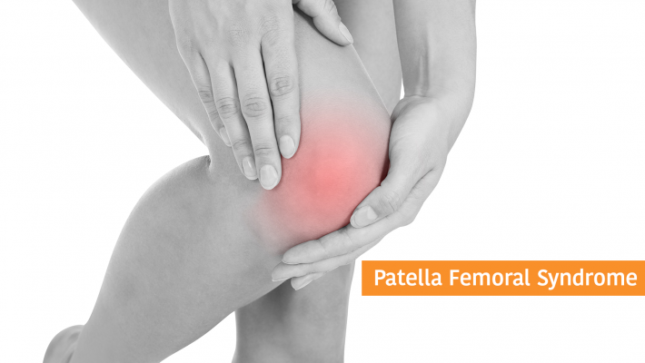
When people think of capsulitis, they tend to think of shoulders and hips but the feet are prone to there fair share of capsulitis too.

So what is capsulitis?

Well simply speaking it is inflammation of the joint capsule. Ligaments around
joints and help form a capsule. Joint capsules help your joints to function properly the
ligaments hold them together. It is these that get inflamed. This can lead to toe dislocation
if it not treated appropriately. In fact, capsulitis is sometimes known as predislocation
syndrome. Capsulitis is a condition that can manifest in people of any age.

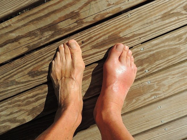
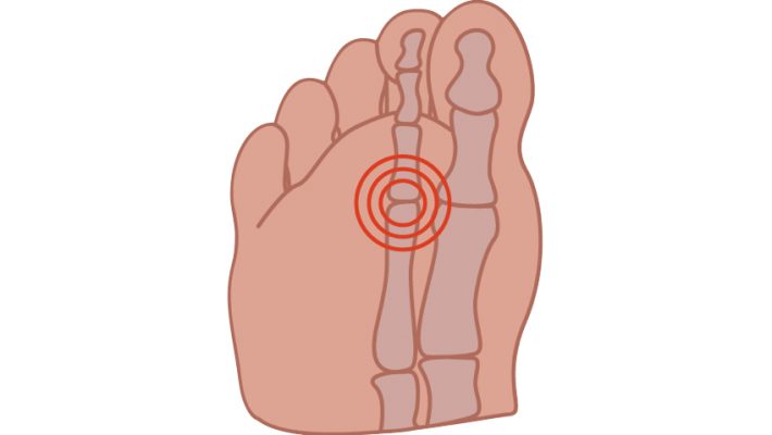
Although any joint in the foot can be subject to capsulatus the 2nd toe joint, under the ball
of the foot is most frequently affected and the metatarsals in general are the joints most
frequently troubled by capsulitis.

Causes vary but increased pressure particularly if the 2nd toe is the longest will cause more
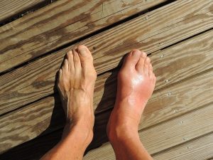
pressure on that metatarsal head. Other causes include large bunions which can also be
prone to capsulitis themselves or by putting more pressure on the adjacent second
metatarsal lead to problems there. An unstable arch of the foot and footwear which may
include high heels, narrow toe box or toe spring which is an elevated toe box common in
many shoes also predispose to this problem. Tight posterior muscle groups and tight or
unbalanced tendons in the foot may further exacerbate the situation. The problem is also
common in runners and sports men and women.

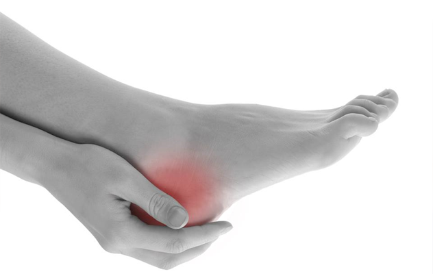
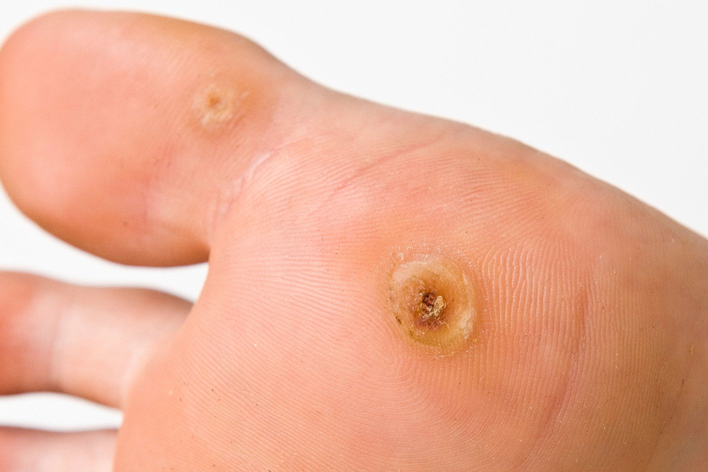
Symptoms, pain is always a feature and other symptoms may include redness, callous over
the area, increased space between toes, the feeling of walking on a stone and swelling
around the area.

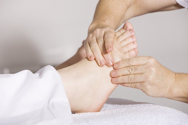
Treatment is directed at the causes, and may include rest and reducing weight bearing
activities, padding, stretching. Insoles are often helpful to deflect the pressure/control the
foot.

Often a cutaway is used to reduce pressure under the area. Icing the area and laser is
very helpful to reduce the inflammation in combination with padding strapping and or
insoles or orthotics.

Corns

Corns

One of the most common problems that I come across in my clinics are corns, so this month I thought I would talk about what they are and more importantly how to deal with them.

Corns are painful lesions on the feet caused by mechanical stress to that particular area. This increased mechanical stress can be due to pressure from foot ware or changes in the foot or the way we walk.

There are several types of corns or Helomata which comes from Greek helos, meaning stone wedge. Hard corns, heloma dura usually occur underneath the foot or on the tops or ends of the toes and may even occur under the nails or in the sides of the nails. Soft corns or heloma molle only appear between the toes and are soft due to perspiration between the toes. Vascular and neurovascular, heloma vasculare and heloma neurovasculare tend to be long standing lesions where blood vessels and nerves have become involved. Finally seed corns or helomata miliare which are not really corns at all. They are not caused by increased stress or pressure on the skin but are simply plugs or beads of cholesterol often in non weight bearing areas.

Differential diagnosis, most commonly confused with verruca which are generally not painful on direct pressure which corns are. Veruca also always have a vascular and neurological element.

Complications, neglected corns are can be prone to infections particularly due to foots close confinement in a warm humid environment and the proximity to the ground. Ulceration may also occur where there is sever mechanical stress or due to loss of sensitivity as in the case of diabetic neuropathy.

Treatment is roughly divided into two parts removal of the corn, sharp debridement where the corn is removed by a podiatrist/chiropodist with a scalpel. This should be a painless procedure and give instant relief from the ongoing discomfort.

Occasionally the use of local anaesthetic is required in the case of neurovascular corns. Once the corn is out the aim of the treatment is to prevent it returning. Here eliminating the original causes is the priority, reducing the pressure from foot ware and mechanical stress.

This may be padding to the foot in the short term. Changing footwear, insoles, orthotics or silicone guards to redistribute pressure where there have been changes in the shape and function of the foot longer term to mitigate the underlying problems.

There are of course over the counter remedies, usually in the form of corn plasters and these come in two basic types.
Foam rings, these aim to reduce the pressure on the lesion. Medicated corn plasters which contain an acid (usually salicylic acid) which aims to soften and dissolve the corn. The danger here is that if great care is not taken medicated corn plasters and liquids can dissolve healthy skin. This may not only be painful but in the case of diabetics and those with poor circulation can also be very dangerous and should be avoided.

As usual my advice is if in doubt see a podiatrist who will be able to treat the presenting problem and also work towards a longer term resolution.
Philip Mann 686912307

Arch Pain

Arch Pain

This month I thought I would write about one of the most common causes of arch pain, Posterior Tibial tendinitis or Posterior Tibial dysfunction as it is sometimes known.

I see this condition a lot in my clinics and it can be a progressively debilitating condition. Which can, if left untreated result in rupture of the tendon.
 
It can present in several ways, a slow progressive onset with pain in the medial side of the arch starting from the middle of the arch. Sometimes the pain will progress around and behind the ankle and up the lower third of the leg. The condition is exacerbated by exercise and even walking can lead to discomfort. Or it can be sudden onset following particularly vigorous exercise or going down particularly hard on the affected foot.
 
Let’s just do a bit of anatomy before we come to the third presentation. Posterior tibial muscle is situated behind the shin bone (Tibia, as the name suggests) towards the outside of the leg. The tendon runs from the lower end of the muscle around the back of the inside ankle (Medial Malleolus) to attach to the Navicular a bone in the middle of the arch. This muscle is to a large extent responsible for pulling the arch of the foot up. So in those people with a low arch this muscle and tendon have to work a lot harder. If the foot flattens out (pronates) then with over use, the individual can experience pain at the attachment of the tendon (ensopathy) in the arch or as the tendon runs behind the ankle bone increased friction and irritation there as I have already mentioned can cause pain around the ankle and into the bottom of the leg.
 
The third presentation is pain on the top of the foot. But why? The attachment is under the arch? Well the body doesn’t like pain so if it hurts to use this tendon to maintain the integrity of the arch we then compensate and use the tendons on the top of the foot to try and do the same thing. Unfortunately they are not designed to do this and start to get inflamed themselves resulting in compensation pain on the top of the foot.
 
Finally there is rupture; this may not be dramatic and sudden as it can slowly progress from partial rupture with increasing lowering of the arch and spreading of the toes away from the midline of the body and turning out of the heel, to total rupture where the arch ends up touching the ground (weight bearing Navicular).
 
Occasionally rupture is sudden often taking the form of a sports injury the patient will feel it go and will require immediate surgical intervention within 24 hours before the tendon retracts up the leg.
 
So what to do? Well there is plenty. Rest and reduce the inflammation, be that with ice, anti-inflammatory drugs or as favoured in my office, Laser to the area. Then supporting the foot either with strapping or an orthotic (arch support)
 
In severe cases, an Air Cast which is a bit like a Ski Boot to immobilise the foot and give the tendon total rest. Usually Posterior Tibial tendinitis is diagnosed from symptoms and palpation, but where rupture or partial rupture is suspected MRI is very useful.
 
Philip Mann Podiatrist / Chiropodist 686 912 307 www.footpodiatrist.com
knee pain

Knee pain and feet

One of the most common knee problems that I see in clinic is Patella Femoral syndrome; also sometimes know as runner’s knee.

The problem causes pain in the front central portion of the knee usually worse when running or walking up or down hill or going up and down stairs (down particularly). Often there can be pain when sitting with knees bent, which is why the condition is also known as theatre goers knee. Pain comes from behind the kneecap (Patella) where it contacts with the thigh bone (femur).

Prolonged excessive shearing forces between the back of the patella and the front of the femur as the patella no longer tracks nicely between the groves formed by the bottom of the femur.

The problem is usually caused by biomechanical dysfunction, over pull of the lateral quadriceps, excessive pronation or supnation (rolling in or rolling out) of the foot. Overuse in sports or activities which are tough on the knee can also lead to patella femoral syndrome, running, jumping, cycling often exacerbated by biomechanical problems.

This results in inflammation, pain and eventually thinning and softening (Chondromalacia) of the articular cartilage behind the patella and on the femur. The condition can also be secondary to fractures, Osteoarthritis and bony tumours, i.e. changes in the bony structure of the knee.

The problem is usually diagnosed on examination and history, although X-ray and MRI can be useful if the diagnosis is not straightforward.

Treatment can involve any or all of the following, controlling excessive biomechanical dysfunction (pronation or supination) of the feet and legs with either strapping or orthotics, quadriceps strengthening, stretching of posterior muscles of the leg and thigh, and sometimes direct treatment to the knee, laser, icing and anti-inflammatory drugs.

Treatment will depend on the severity and duration of the problem and identification of the underlying causes. Prognosis is full recovery, and a return to normal activities once underlying causes are identified and addressed. I think podiatrists are in a unique position the treat this problem as we are able to address the underlying biomechanical problems to resolve the condition. But then I am a bit biased!

Philip Mann Podiatrist/Chiropodist tel: 686 912 307 or www.footpodiatrist.com

Achilles Tendinitis

Achilles Tendinitis

I seem to be seeing a lot of Achilles Tendinitis in the clinics at the moment. Also known as Achilles tenosynovitis or Achilles tendinopathy.

This condition causes pain in the back of the heel, worse on and after exercise and often particularly painful on rising in the morning. There may be swelling and thickening of the tendon, it may look like a nodule on the back of the tendon.

There are two main presentations of this condition, Noninsertional Achilles Tendinitis and Insertional Achilles Tendinitis, as the names suggest, insertional is in the part of the tendon which attaches into the bone and noninsertional is in the other part. Sometimes the insertional variety is associated with a bony spur formation at the back of the heel (not to be confused with calcaneal heel spur).

So what causes Achilles Tendinitis?

Well as usual there a number of causes it tends to be an overuse type injury, although tight calf muscle in my experience is always present.

Sudden increase in exercise duration or intensity, insufficient stretching before and after exercise, worn shoes and
over pronation where the foot flattens can all contribute.

The condition is common in runners and also sports which involve jumping but can also affect people who are not sporty at all.

The achilles tendon is the largest tendon in the body and it attaches the calf muscle to the back of the heel. It is used to lift the heel and it stores energy each step to help with the following step although very strong it has to have some elasticity.

When someone gets Achilles Tendinitis there is scarring and degradation of the tendon which causes the tendon to become stiff and inflexible. This increases the possibility of rupture. Due to the poor blood supply to achilles it can take a long time to heal. The literature often says six to twelve months although with good treatment I expect it to be a lot quicker.

Treatment of Achilles Tendinitis involves stretching and strengthening, I find Low level Laser works very well as it increases the blood supply to the area and also reduces the pain and swelling.

Rest and cessation of activities which exacerbate the problem, switching to low impact exercise. Ice and anti inflammatories help.

Addressing footwear and controlling the over pronation with strapping or and orthotic. I tend to favor orthotics with a combined heel raise which control pronation and reduce the velocity of the pull on achilles. In severe cases surgical debridement of the tendon may be required.

As with all overuse/sports type injuries there is no single cause to the problem and therefore no single solution and successful treatment is tailored to the individual’s causes and needs.

Philip Mann Podiatrist/Chiropodist 686912307 www.footpodiatrist.com

Permanent Solutions to Ingrown Toenails

A PERMANENT SOLUTION TO INGROWN TOENAILS

Ingrown toenails are one of the most painful conditions that I treat in my clinics, but the treatment itself is virtually painless.

It always surprises me when meet patients who have been having an ingrown toenail treated for a long time sometimes many years and they have not been offered a permanent solution to the problem.

It makes me suspicious that either the person who is treating them wants them to keep coming back to have the same problem treated, or that they are not qualified to do the surgery and therefore working illegally.

In order to work in Spain as a podiatrist/chiropodist you must be able to to do basic surgery under local anesthetic. But I am getting a bit ahead of myself.

What is an ingrown toenail?

Well it’s where part of the edge or front of the nail pushes into the skin causing the toe to become sore swollen and inflamed. The condition occurs mainly on the 1st toe and is really very much like having a splinter. It is a foreign body in the skin which needs removing. Just like a splinter once removed the pain disappears and the area settles down and returns to normal.

Sometimes that is all that is needed, if the nail was cut badly taking away that sharp or rough edge is all that is required a little careful cutting by someone who knows what they are doing and is at the right end of the body to see what they are doing.

Unfortunately often the problem re occurs this is usually because the nail has become a little curved. This is when a permanent solution should at least be offered.

This is how it works in my clinics, if you present with an ingrown toenail, initially I will remove the bit and make you comfortable on the first visit, I will talk about surgery as an option but this option is up to the patient. My advice is usually something like. If this a problem once or twice a year come back and I will just cut a little bit away.

But if this keeps being a problem every month or two then you should consider surgery as it is a permanent solution. This is elective surgery so you decide if you want it and when it is convenient to have the procedure. I am happy to keep cutting a little bit off if that is what the patient wants. Some people are happy with that others may not be suitable for surgery due to problems with health, circulation and diabetes are considerations.

Ninety percent of the time I do not remove the whole nail but just a strip all the way down the side so after you still have a nail and it doesn’t really look much different except I have removed the bit that curved into the skin.

Although I do not like to say it is a totally painless procedure, the vast majority of people and I do a lot of this on children, say it is virtually pain free. The anesthetic is put in at the base of the toe where it joins the foot away from the sore bit and this makes the whole toe numb. Once the part of nail is removed the area is treated with phenol to prevent the nail growing back. I allow people to swim, shower and get on with life following the surgery but most people will want to be in an open shoe for at least a week following the procedure.

If you have been suffering for a long time it is really something to consider and nothing to fear.

Philip Mann Podiatrist Chiropodist 686 912 307

Heel pain

Heel Pain

Heel pain is one of the most common problems that I see in the clinic and can be a very debilitating condition

Probably the most common type of heel pain is Plantar Fasciitis or as it is sometimes known as Calcaneal heel spur syndrome. Although heel spurs are often visible on X-ray this is not always diagnostic as they are also often seen on the non symptomatic foot too, and diagnosis is usually on history and symptoms. This condition is characterized by increased pain with the first steps in the morning or when standing after a prolonged period of sitting. It can be in one or both feet. There are many treatments available for this condition depending on the severity of the condition the length of time the individual has had the problem and level of activity, and can vary from stretching and strapping to orthotics and even night splints.

Achilles tendonitis is usually a sports injury or can be caused by over use or even foot ware. Pain is often worse on rising in the morning and exacerbated by activity. Treatment, laser treatment is very effective here and stretching is important also altering foot ware can also help.

Hagland’s deformity or Pump bumps as they are sometimes known are boney lumps at the back of the heel on the outside. Caused by the foot rubbing on foot ware the body attempts to protect the area laying down more bone. Sometimes there is an associated fluid filled sac called a bursa associated with this condition. The area can become inflamed and very sore in shoes. Treatment, controlling the motion of the foot, padding and laser work well, although surgical intervention is sometimes called for.

Bursa can also be a problem behind the Achilles tendon and in the fat pad of the heel. If these bursa become inflamed they can be painful on activity. Once again laser can be very good here as can padding or an accommodating insole.

Sever’s disease is a heel condition only seen in children and adolescents, eight to about 16 years old and is quite common. Usually seen in very sporty individuals, pain is exacerbated by exercise and often is only present when running or jumping. Often Sever’s coincides with a grown spurt as the leg bone grows and the Achilles tendon pulls on the heel bone pulling the growth plate of the bone away and causing localized avascular necrosis with resultant pain on exercise. Orthotics and stretching work very well here enabling the child recover fully and return to the sports they enjoy.

There are many conditions which cause heel pain from tumors, fractures to simple splits. I have just covered a few examples here.

Diabetic foot care

Why Diabetic foot care is important

With the incidence of diabetes on the increase in the western world, the importance of foot care for diabetics has never been more important. Sadly diabetics represent the biggest group of lower limb amputees even greater than that of road traffic accidents. But it is not all bad news diabetic amputations are avoidable and in this article we will look at the causes and how to avoid problems leading to amputation.

Diabetes is an increase in circulating blood sugar levels; there are two types of Diabetes, Type I and Type II. Type I often known as juvenile diabetes the onset is in childhood and young adults Type II diabetes, usually known as mature onset diabetes, as the name suggests mainly starts later in life. Both diseases are controlled differently, Type I requiring injections of insulin and Type II may be controlled by reducing sugar intake (diet) or drugs which help the body to use insulin better. Type II may eventually require the use or insulin too. Ultimately the disease causes the same problems to the body, especially if uncontrolled or poorly controlled. There is damage to small blood vessels particularly in feet, hands, eyes, kidneys and heart and damage to nerves.

Manifestations in the foot are largely three fold, poor circulation, nerve damage and poor healing following injury. But just because you are diabetic it does not mean that you will automatically have all of the following problems. If diabetes is well controlled it has a minimal effect on the body so controlling your blood sugar levels is very important.

Circulation can be effected by accelerating arthrosclerosis, the hardening of the arteries; this makes it more difficult for the blood to get to where it needs to go. If the blood can’t get down there in sufficient quantity or quality, when an injury occurs it may be slow to heal or even ulcerate. If the ulceration is not treated it can get infected and if that infection is not treated it can turn to gangrene which would mean amputation of the affected toe, foot or even leg. But as you can see there are many chances for intervention, from treating the original injury to treating the ulceration to treating the infection. Obviously it is better to treat the original injury early and avoid the complications of ulceration and infection.

Nerve damage or neuropathy can occur with diabetes. This can lead to loss of feeling particularly pain sensation in the feet. The problem here is the individual may not be aware that there is loss of feeling and if there is an injury may not seek care promptly. This increases the dangers of the above complications. Over the years my diabetic patients have been extremely inventive when it comes to damaging their feet without realizing it. From simple things like blisters and sun burn to scalding feet in a hot bath to the extreme cases of walking round with a nail through their shoe into the foot, all treatable but also all avoidable.

Poor healing in diabetic feet means that injuries are often slow to heal so wounds stay open longer increasing the chances of getting infected and infections love sugar. The higher the blood sugar the more rapidly the infection can take hold.

So what can we do to avoid these problems? Well, plenty. Good diabetic control means less damage and less problems. Inspect the feet daily make sure there are no injuries or changes in colour, inflammation or pain, if need be look at the bottom of the feet in a mirror if you have problems getting down there. Don’t walk bare foot it’s just asking for trouble. Wash the feet daily and dry carefully between toes and apply moisturiser to any dry areas, but not between the toes. Have feet measured when buying shoes and always check inside shoe before putting it on for stones or other foreign bodies. Visit a Podiatrist, well I would say that, but foot assessment by a podiatrist is important, circulation can be checked with Doppler and neuropathy measured then tailored advice given as well as routine care of nails corns and callous and even ulcers. If you have a foot problem don’t ignore it or try to self treat or self diagnose. Unfortunately there are too many amputees who put off seeking medical attention worried as they had heard about diabetics losing limbs, so that by the time they actually do seek care it is too late. Early intervention and treatment is the key to avoiding serious foot problems and having peace of mind.

Unwelcome visitors

I am not talking about the ones who use the last of the milk or think that a fresh towel is required for every trip to the shower these ones are far worse. Verrucas are the subject this month. So what are they? how do we get them? and more importantly how do we get rid of them?

Verruca pedis, to give them their full name, or ‘plantar warts’ as they are also sometimes known. are a benign (non cancerous) viral infection of the skin most commonly found on the soles of the feet. The infection is caused by the human papilloma virus (HPV). The virus can cause different types of verruca, from single lesions to multiple clusters known as mosaic lesions and can also be spread to other parts of the body. Verruca occur most often in children and young adults, between the ages of 12 and 16 but adults can also be affected. Incidence is higher in people who share common bathing areas (e.g., dormitory students, gym members). This may well be why I see more verruca here as we have so many swimming pools.

Verrucas can occur when HPV invades the body through tiny cuts or breaks in the skin on the bottom of the feet. The virus often is encountered on contaminated surfaces, such as the tile floors of public changing rooms, showers, and swimming pools. Normally, antibodies in the blood destroy HPV, but in some cases, it takes refuge in the skin and verruca appear

Appearance and diagnosis. Usually spongy in appearance often grey or yellowish with a well defined border. Small back dots may be seen within the lesions, these are capillaries which supply them with blood. They are often likened to icebergs because the part that you see on the surface is often half the size of what is underneath. They may be painful as they may occur under areas of pressure on the feet, like the ball of the foot or the heel. Generally verruca are diagnosed on appearance and by the “squeeze test”, verruca are not usually painful on direct pressure, unlike corns and callous, but squeezing either side of the lesion causes sharp pain. A word of warning to parents wanting to try this test on their children , squeeze very gently at first as a small amount of pressure can cause a large amount of pain. Differential diagnoses are most commonly corns or callous but in rare circumstances other benign and even malignant lesions have to be considered. Occasionally if there is doubt over the diagnosis a skin scraping may be taken by the podiatrist for analysis by the pathology lab.

So how do we treat these unwelcome guests? Well there are plenty of home treatments available over the counter at the pharmacy. These are usually in the form of acids, salicylic acid being the most common. Care needs to be taken not to damage uninfected skin and these treatments should be avoided if the patient is diabetic or sufferers from circulatory problems. I have also come across a number of rather unconventional treatments over the years. Duct tape has its supporters and there was some research which indicated that it was as effective as many of the over the counter medicines. But this may reflect how ineffective some over the counter medicines are rather than the effectiveness of duct tape. Garlic applied to the lesion daily is also said to be effective, I had a fellow podiatrist in Bermuda who used to swear by it, although I am yet to be convinced. Homeopathy in the form of marigold is also said to be effective.

If home remedies prove ineffective, then a trip to someone like me may be required. A podiatrist will have a number of treatments available to treat verruca. Topical chemical treatments similar to home treatments but a lot stronger combined with debridement enable better penetration of the infected tissue. These topical chemical treatments will need to be repeated over a number weeks to completely kill the verruca. More aggressive and often more effective is cryosurgery. Here the infected tissue is frozen with liquid nitrogen or similar cryogen. Freezing damages the cells causing cell death end eventually resolution. Unfortunately there is no one hundred percent effective treatment for all verruca and although most lesions only require a little help from the podiatrist occasionally we do come across very stubborn ones which require multiple treatments over an extended period and patience and persistence are required on the part of the patient and the practitioner. So treatment can be tricky, what can we do to avoid them? Well here’s what our friends at the American Podiatric Medical Association (APMA) have to say, the following may help to prevent plantar warts: (verruca) Avoid walking barefoot whenever possible.

  • Change shoes and socks daily.
  • Keep feet clean and dry.
  • Check children’s feet periodically.
  • Avoid direct contact with warts on other persons or on other parts of the body.
  • Do not ignore growths on, or changes in, your skin.
  • Visit a podiatrist as part of your annual health check-up.

Foot Pain

As the interface between the body and the ground the foot is subject to enormous stresses and loads during normal walking and running. With the hard unyielding ground and the rotations of the leg and trunk acting on it this complex structure it’s not surprising that occasionally we have problems. Sports men and women are more prone to problems as they demand more from their feet and legs.

When, we walk the first part of the foot to hit the ground is the outside of our heel (this is why we wear this part of our shoe first); as the rest of the foot comes in contact with the ground we start to move our body weight over the foot. At this point the foot starts to pronate (the rolling and flattening of the arch) this is a normal part of the gait cycle and allows the foot to adapt to uneven surfaces and absorb shock. As the body continues to move over the foot the foot starts to supernate, the arch starts to rise and the foot becomes more of a rigid lever enabling us to pivot over the foot (or push off) so we can take our next step, and so the cycle continues. We take an average of 2500 steps a day which means we walk about 3000 miles a year. It is not difficult to imagine that small changes in the gait cycle could have a significant impact on the whole system. If for example there is excessive pronation or flattening of the arch during the gait cycle, then we may be trying to pivot (push off) a foot that is a mobile adaptor rather than a rigid lever. Not only would this be less effective at moving us forward and therefore require more effort, but it may also have an impact on the structure doing the moving, i.e. the feet and legs. Excessive pronation can lead to many foot and leg conditions including, plantar fasciitis, tendonitis, shin splints, patello femoral problems and capsulitus to name a few. There may well be more remote symptoms including back pain and even tempro mandibular joint problems (jaw pain). Equally if there is excessive supernation and the foot is a rigid lever when it should be a mobile adaptor then the feet will be poor shock absorbers and this may also lead to problems like ankle sprains, Morton’s neuroma, stress fractures and knee problems, once again to name a few. Even callous and corns can often be traced back to underlying biomechanical problems causing increased pressure and or friction on a certain part of the foot.

Part of the role of a podiatrist is to assess the patient’s biomechanical problems when appropriate. This usually takes the form of a biomechanical assessment. The patient is examined to see how everything lines up, feet, legs, knees, and the rest of the body, lines may be drawn on the legs and feet and ranges of joint motion measured. Weakness or tightness of associated muscles will be noted. The gait may be observed and foot ware examined. From this the podiatrist can then make a diagnosis and prescribe the appropriate care often in the form of a custom made orthotic which fits in the shoe, this reduces excessive pronation or supernation and helps to align the legs and feet. Muscle stretching or strengthening may also be required. Often direct treatment to the injury or problem could be needed in the form of manipulation, strapping or laser treatment to the area. This type of treatment can not only resolve the presenting problem but also improve the way an individual walks or runs helping them to go faster or longer and preventing further injuries. Over the years I have treated many sports men and women from ramblers and power walkers, junior tri athletes, to professional footballers and marathon runners.