Charcot Foot Example

Charcot Foot

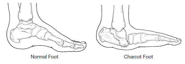
Charcot foot is a condition associated with neuropathy (nerve damage) to the foot.

It can lead to deformity, ulceration and even amputation. Although this problem is associated with many conditions that involve neuropathy to the feet, these include nerve damage caused by toxins (ethanol, drug related), infection (leprosy), as well as spinal cord damage and a number of other diseases (Parkinson’s disease, HIV, sarcoidosis, rheumatoid arthritis and psoriasis).

But by far the most common underlying problem is Diabetes, in over 20 years of looking at feet I have only seen one patient with a Charcot foot who was not diabetic.

There is still some, shall we say discussion in the medical profession as to the exact disease process, but most agree that weakened bones in the foot exacerbated by loss of feeling and sometimes trauma start off an inflammatory process. The current belief is that once the disease is triggered in a susceptible individual, there is uncontrolled inflammation in the foot. This inflammation leads to bone breaking down and is indirectly responsible for the progressive fracture and dislocation of joints in the foot.

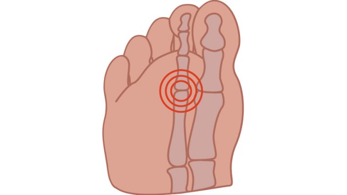
Charcot foot symptoms, always include swelling of the foot often without a history of trauma. In the early stages there may be redness and warmth and sometimes pain. Although often diagnosed by examination and patient history x-ray is useful to confirm diagnosis. Changes to the bone that are seen on x-ray may be confused for a bone infection. A bone infection is very unlikely if the skin is intact and there is no ulcer present.

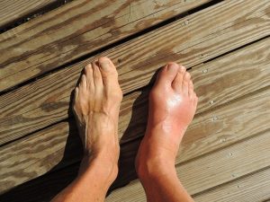
Treatment involves immobilising the foot usually with a cast or boot until the inflammation has stabilised. A Charcot episode usually results in changes to the structure of the foot often collapsing the arch of the foot resulting in a ‘rocker’ type foot. Following a Charcot episode an insole to support the foot and prevent further damage and charcot changes is required.

Contact me if you have any questions or would like further advice

Capsulitis feet

Capsulitis and feet.

When people think of capsulitis, they tend to think of shoulders and hips but the feet are prone to there fair share of capsulitis too.

So what is capsulitis?

Well simply speaking it is inflammation of the joint capsule. Ligaments around
joints and help form a capsule. Joint capsules help your joints to function properly the
ligaments hold them together. It is these that get inflamed. This can lead to toe dislocation
if it not treated appropriately. In fact, capsulitis is sometimes known as predislocation
syndrome. Capsulitis is a condition that can manifest in people of any age.

Although any joint in the foot can be subject to capsulatus the 2nd toe joint, under the ball
of the foot is most frequently affected and the metatarsals in general are the joints most
frequently troubled by capsulitis.

Causes vary but increased pressure particularly if the 2nd toe is the longest will cause more
pressure on that metatarsal head. Other causes include large bunions which can also be
prone to capsulitis themselves or by putting more pressure on the adjacent second
metatarsal lead to problems there. An unstable arch of the foot and footwear which may
include high heels, narrow toe box or toe spring which is an elevated toe box common in
many shoes also predispose to this problem. Tight posterior muscle groups and tight or
unbalanced tendons in the foot may further exacerbate the situation. The problem is also
common in runners and sports men and women.

Symptoms, pain is always a feature and other symptoms may include redness, callous over
the area, increased space between toes, the feeling of walking on a stone and swelling
around the area.

Treatment is directed at the causes, and may include rest and reducing weight bearing
activities, padding, stretching. Insoles are often helpful to deflect the pressure/control the
foot.

Often a cutaway is used to reduce pressure under the area. Icing the area and laser is
very helpful to reduce the inflammation in combination with padding strapping and or
insoles or orthotics.

Corns

Corns

One of the most common problems that I come across in my clinics are corns, so this month I thought I would talk about what they are and more importantly how to deal with them.

Corns are painful lesions on the feet caused by mechanical stress to that particular area. This increased mechanical stress can be due to pressure from foot ware or changes in the foot or the way we walk.

There are several types of corns or Helomata which comes from Greek helos, meaning stone wedge. Hard corns, heloma dura usually occur underneath the foot or on the tops or ends of the toes and may even occur under the nails or in the sides of the nails. Soft corns or heloma molle only appear between the toes and are soft due to perspiration between the toes. Vascular and neurovascular, heloma vasculare and heloma neurovasculare tend to be long standing lesions where blood vessels and nerves have become involved. Finally seed corns or helomata miliare which are not really corns at all. They are not caused by increased stress or pressure on the skin but are simply plugs or beads of cholesterol often in non weight bearing areas.

Differential diagnosis, most commonly confused with verruca which are generally not painful on direct pressure which corns are. Veruca also always have a vascular and neurological element.

Complications, neglected corns are can be prone to infections particularly due to foots close confinement in a warm humid environment and the proximity to the ground. Ulceration may also occur where there is sever mechanical stress or due to loss of sensitivity as in the case of diabetic neuropathy.

Treatment is roughly divided into two parts removal of the corn, sharp debridement where the corn is removed by a podiatrist/chiropodist with a scalpel. This should be a painless procedure and give instant relief from the ongoing discomfort.

Occasionally the use of local anaesthetic is required in the case of neurovascular corns. Once the corn is out the aim of the treatment is to prevent it returning. Here eliminating the original causes is the priority, reducing the pressure from foot ware and mechanical stress.

This may be padding to the foot in the short term. Changing footwear, insoles, orthotics or silicone guards to redistribute pressure where there have been changes in the shape and function of the foot longer term to mitigate the underlying problems.

There are of course over the counter remedies, usually in the form of corn plasters and these come in two basic types.
Foam rings, these aim to reduce the pressure on the lesion. Medicated corn plasters which contain an acid (usually salicylic acid) which aims to soften and dissolve the corn. The danger here is that if great care is not taken medicated corn plasters and liquids can dissolve healthy skin. This may not only be painful but in the case of diabetics and those with poor circulation can also be very dangerous and should be avoided.

As usual my advice is if in doubt see a podiatrist who will be able to treat the presenting problem and also work towards a longer term resolution.
Philip Mann 686912307

Arch Pain

Arch Pain

This month I thought I would write about one of the most common causes of arch pain, Posterior Tibial tendinitis or Posterior Tibial dysfunction as it is sometimes known.

I see this condition a lot in my clinics and it can be a progressively debilitating condition. Which can, if left untreated result in rupture of the tendon.
 
It can present in several ways, a slow progressive onset with pain in the medial side of the arch starting from the middle of the arch. Sometimes the pain will progress around and behind the ankle and up the lower third of the leg. The condition is exacerbated by exercise and even walking can lead to discomfort. Or it can be sudden onset following particularly vigorous exercise or going down particularly hard on the affected foot.
 
Let’s just do a bit of anatomy before we come to the third presentation. Posterior tibial muscle is situated behind the shin bone (Tibia, as the name suggests) towards the outside of the leg. The tendon runs from the lower end of the muscle around the back of the inside ankle (Medial Malleolus) to attach to the Navicular a bone in the middle of the arch. This muscle is to a large extent responsible for pulling the arch of the foot up. So in those people with a low arch this muscle and tendon have to work a lot harder. If the foot flattens out (pronates) then with over use, the individual can experience pain at the attachment of the tendon (ensopathy) in the arch or as the tendon runs behind the ankle bone increased friction and irritation there as I have already mentioned can cause pain around the ankle and into the bottom of the leg.
 
The third presentation is pain on the top of the foot. But why? The attachment is under the arch? Well the body doesn’t like pain so if it hurts to use this tendon to maintain the integrity of the arch we then compensate and use the tendons on the top of the foot to try and do the same thing. Unfortunately they are not designed to do this and start to get inflamed themselves resulting in compensation pain on the top of the foot.
 
Finally there is rupture; this may not be dramatic and sudden as it can slowly progress from partial rupture with increasing lowering of the arch and spreading of the toes away from the midline of the body and turning out of the heel, to total rupture where the arch ends up touching the ground (weight bearing Navicular).
 
Occasionally rupture is sudden often taking the form of a sports injury the patient will feel it go and will require immediate surgical intervention within 24 hours before the tendon retracts up the leg.
 
So what to do? Well there is plenty. Rest and reduce the inflammation, be that with ice, anti-inflammatory drugs or as favoured in my office, Laser to the area. Then supporting the foot either with strapping or an orthotic (arch support)
 
In severe cases, an Air Cast which is a bit like a Ski Boot to immobilise the foot and give the tendon total rest. Usually Posterior Tibial tendinitis is diagnosed from symptoms and palpation, but where rupture or partial rupture is suspected MRI is very useful.
 
Philip Mann Podiatrist / Chiropodist 686 912 307 www.footpodiatrist.com
knee pain

Knee pain and feet

One of the most common knee problems that I see in clinic is Patella Femoral syndrome; also sometimes know as runner’s knee.

The problem causes pain in the front central portion of the knee usually worse when running or walking up or down hill or going up and down stairs (down particularly). Often there can be pain when sitting with knees bent, which is why the condition is also known as theatre goers knee. Pain comes from behind the kneecap (Patella) where it contacts with the thigh bone (femur).

Prolonged excessive shearing forces between the back of the patella and the front of the femur as the patella no longer tracks nicely between the groves formed by the bottom of the femur.

The problem is usually caused by biomechanical dysfunction, over pull of the lateral quadriceps, excessive pronation or supnation (rolling in or rolling out) of the foot. Overuse in sports or activities which are tough on the knee can also lead to patella femoral syndrome, running, jumping, cycling often exacerbated by biomechanical problems.

This results in inflammation, pain and eventually thinning and softening (Chondromalacia) of the articular cartilage behind the patella and on the femur. The condition can also be secondary to fractures, Osteoarthritis and bony tumours, i.e. changes in the bony structure of the knee.

The problem is usually diagnosed on examination and history, although X-ray and MRI can be useful if the diagnosis is not straightforward.

Treatment can involve any or all of the following, controlling excessive biomechanical dysfunction (pronation or supination) of the feet and legs with either strapping or orthotics, quadriceps strengthening, stretching of posterior muscles of the leg and thigh, and sometimes direct treatment to the knee, laser, icing and anti-inflammatory drugs.

Treatment will depend on the severity and duration of the problem and identification of the underlying causes. Prognosis is full recovery, and a return to normal activities once underlying causes are identified and addressed. I think podiatrists are in a unique position the treat this problem as we are able to address the underlying biomechanical problems to resolve the condition. But then I am a bit biased!

Philip Mann Podiatrist/Chiropodist tel: 686 912 307 or www.footpodiatrist.com

Achilles Tendinitis

Achilles Tendinitis

I seem to be seeing a lot of Achilles Tendinitis in the clinics at the moment. Also known as Achilles tenosynovitis or Achilles tendinopathy.

This condition causes pain in the back of the heel, worse on and after exercise and often particularly painful on rising in the morning. There may be swelling and thickening of the tendon, it may look like a nodule on the back of the tendon.

There are two main presentations of this condition, Noninsertional Achilles Tendinitis and Insertional Achilles Tendinitis, as the names suggest, insertional is in the part of the tendon which attaches into the bone and noninsertional is in the other part. Sometimes the insertional variety is associated with a bony spur formation at the back of the heel (not to be confused with calcaneal heel spur).

So what causes Achilles Tendinitis?

Well as usual there a number of causes it tends to be an overuse type injury, although tight calf muscle in my experience is always present.

Sudden increase in exercise duration or intensity, insufficient stretching before and after exercise, worn shoes and
over pronation where the foot flattens can all contribute.

The condition is common in runners and also sports which involve jumping but can also affect people who are not sporty at all.

The achilles tendon is the largest tendon in the body and it attaches the calf muscle to the back of the heel. It is used to lift the heel and it stores energy each step to help with the following step although very strong it has to have some elasticity.

When someone gets Achilles Tendinitis there is scarring and degradation of the tendon which causes the tendon to become stiff and inflexible. This increases the possibility of rupture. Due to the poor blood supply to achilles it can take a long time to heal. The literature often says six to twelve months although with good treatment I expect it to be a lot quicker.

Treatment of Achilles Tendinitis involves stretching and strengthening, I find Low level Laser works very well as it increases the blood supply to the area and also reduces the pain and swelling.

Rest and cessation of activities which exacerbate the problem, switching to low impact exercise. Ice and anti inflammatories help.

Addressing footwear and controlling the over pronation with strapping or and orthotic. I tend to favor orthotics with a combined heel raise which control pronation and reduce the velocity of the pull on achilles. In severe cases surgical debridement of the tendon may be required.

As with all overuse/sports type injuries there is no single cause to the problem and therefore no single solution and successful treatment is tailored to the individual’s causes and needs.

Philip Mann Podiatrist/Chiropodist 686912307 www.footpodiatrist.com

Dermatology and Feet

Dermatology and Feet

One of the many facets of podiatry is dermatology and if you think about it, feet are covered in skin and nails are just an adaption of skin.

There is now a podiatric speciality known as Podiatric dermatology which has grown out of the fact, that as a podiatrist much of what you do crosses over into dermatology and therefore many podiatrists want to know even more and specialise in this area.

When I go to conferences dermatology lectures are always packed. Many of the routine treatments I am involved in are dermatological in nature, Hyperkeratosis (callous), Heloma durum (corns), Human papiloma virus (verrucas) Onycomycosis (nail fungus) are all dermatological complaints which podiatrists treat every day. There are also the more serious skin conditions that often present in clinic.

Skin cancer is very common in feet as after to hands and faces feet are exposed to the sun probably more than any part of the body. From solar Keratosis sometimes known as sun spots, Squamous cell carcinoma, Basal cell carcinoma and even malignant Melanoma are all commonly seen on feet.

I think I have mentioned before that one of the differential diagnosis for ingrown toenails particularly if they are persistent, is malignant Melanoma. Obviously with skin cancers it is not a podiatrist’s role to treat them and swift referral especially in the case of suspected Melanoma to a Dermatologist is essential.

But malignancy aside, there are still many skin conditions of the feet that podiatrists treat very well. Cracked heels are very treatable with debridement and advice on the correct emollients. Often cracked or split heels are a symptom of psoriasis and psoriasis has other manifestations in the feet one of which is psoriatic nails. These often look just like fungal nails and although the psoriasis in nails cannot be eliminated it can be managed very well by reducing the nail bulk and improving appearance.

Athlete’s foot which is really a fungal infection is a very common presentation in clinic in its many forms. Even bacterial infections like Pitted Keratolysis where white holes are seen in the bottom of the feet especially when wet are very treatable in clinic.

There are too many skin conditions seen in feet to mention them all, but the above are a few which are commonly seen.

Permanent Solutions to Ingrown Toenails

A PERMANENT SOLUTION TO INGROWN TOENAILS

Ingrown toenails are one of the most painful conditions that I treat in my clinics, but the treatment itself is virtually painless.

It always surprises me when meet patients who have been having an ingrown toenail treated for a long time sometimes many years and they have not been offered a permanent solution to the problem.

It makes me suspicious that either the person who is treating them wants them to keep coming back to have the same problem treated, or that they are not qualified to do the surgery and therefore working illegally.

In order to work in Spain as a podiatrist/chiropodist you must be able to to do basic surgery under local anesthetic. But I am getting a bit ahead of myself.

What is an ingrown toenail?

Well it’s where part of the edge or front of the nail pushes into the skin causing the toe to become sore swollen and inflamed. The condition occurs mainly on the 1st toe and is really very much like having a splinter. It is a foreign body in the skin which needs removing. Just like a splinter once removed the pain disappears and the area settles down and returns to normal.

Sometimes that is all that is needed, if the nail was cut badly taking away that sharp or rough edge is all that is required a little careful cutting by someone who knows what they are doing and is at the right end of the body to see what they are doing.

Unfortunately often the problem re occurs this is usually because the nail has become a little curved. This is when a permanent solution should at least be offered.

This is how it works in my clinics, if you present with an ingrown toenail, initially I will remove the bit and make you comfortable on the first visit, I will talk about surgery as an option but this option is up to the patient. My advice is usually something like. If this a problem once or twice a year come back and I will just cut a little bit away.

But if this keeps being a problem every month or two then you should consider surgery as it is a permanent solution. This is elective surgery so you decide if you want it and when it is convenient to have the procedure. I am happy to keep cutting a little bit off if that is what the patient wants. Some people are happy with that others may not be suitable for surgery due to problems with health, circulation and diabetes are considerations.

Ninety percent of the time I do not remove the whole nail but just a strip all the way down the side so after you still have a nail and it doesn’t really look much different except I have removed the bit that curved into the skin.

Although I do not like to say it is a totally painless procedure, the vast majority of people and I do a lot of this on children, say it is virtually pain free. The anesthetic is put in at the base of the toe where it joins the foot away from the sore bit and this makes the whole toe numb. Once the part of nail is removed the area is treated with phenol to prevent the nail growing back. I allow people to swim, shower and get on with life following the surgery but most people will want to be in an open shoe for at least a week following the procedure.

If you have been suffering for a long time it is really something to consider and nothing to fear.

Philip Mann Podiatrist Chiropodist 686 912 307

who would be a podiatrist?

Who would be a Podiatrist?

Without doubt the question I get asked most at work is why would you want to be a podiatrist and look at smelly feet all day?

The question is often a lightly veiled way of saying do you have a foot fetish or something and some people even ask this outright.

This is usually followed by: Have you always wanted to do this since you were at school?

This normally happens about half way through the first consultation. They have already told me what the nature of the problem is I have taken a medical history and asked them questions about their symptoms and I am probably doing some kind of treatment.

I suppose the question is fair enough; on the face of it looking at feet all day is not the most glamorous way to spend your day.

Well in reverse order this is what I normally tell people. I haven’t always wanted to do this. When I left school I trained as a dental technician and made false teeth until I was about in my mid twenty’s.

No I do not have a foot fetish. As to why? Well I really enjoy my work, most people I see come to me in pain and go away feeling better. So there is lots of job satisfaction and people are usually pleased to see me.

There is also a tremendous amount of variation in what I do each day, from nail cutting and corns to looking at people’s biomechanics and gait analysis, minor surgery under local anaesthetic for removal of part of or a whole toe nails, assessment and treatment of diabetic foot problems, wound care, children’s foot problems, sports injuries, treatment of verrucas to name but a few.

Also there is quite a lot of problem solving in my work, clients will come in with a pain in their foot there may be very little to see in the way of obvious signs like swelling and redness and I have to diagnose and treat the problem.

Sometimes a treatment that works for one individual will not be as successful for another with the same condition so an alternative solution tailored to the individual must be found. This all makes my work interesting and rewarding.

Then of course I get to spend at least half an hour a time with my patients and we get to chat, so I get to know people and people are generally interesting. And I know I am not alone, most of my collages are enthusiastic and passionate about what they do. If I go to a conference podiatrists are always keen to learn new techniques and share experience and knowledge.

Don’t get me wrong I don’t get up every morning and leap out of bed and go” great feet again today!” but I do consider myself very lucky to have a job I really enjoy.